Fig. 3.

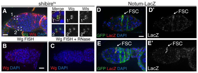
Wg is expressed in escort cells and Wg signaling is active specifically in FSCs. (A) A germarium from a shibirets fly that was shifted to the non-permissive temperature for 2 hours prior to dissection stained for Wg (green), Wntless (red) and with DAPI (blue). Wg+ puncta are visible throughout the region containing escort cells and many colocalize with Wntless. Boxed regions 1 and 2 are magnified to the right and shown as a merged image (1,2), Wg channel only (1′,2′) and Wntless channel only (1′,2′). The dashed line in box 1 shows a nucleus of a shape and position characteristic of escort cells. (B,C) Wild-type germaria stained with a FISH probe for wg transcript (red) and DAPI. Pretreatment of tissue with RNase (C) eliminates the signal, demonstrating that the FISH probe is specific for an RNA target. (D,E) The Wg pathway activity reporter Notum-lacZ is expressed in the anteriormost labeled cell of a GFP+ FSC clone. Tissue is stained for GFP (green), lacZ (β-galactosidase, red) and with DAPI. (D′,E′) The lacZ channel only. Anterior is to the left. Scale bars: 5 μm.
