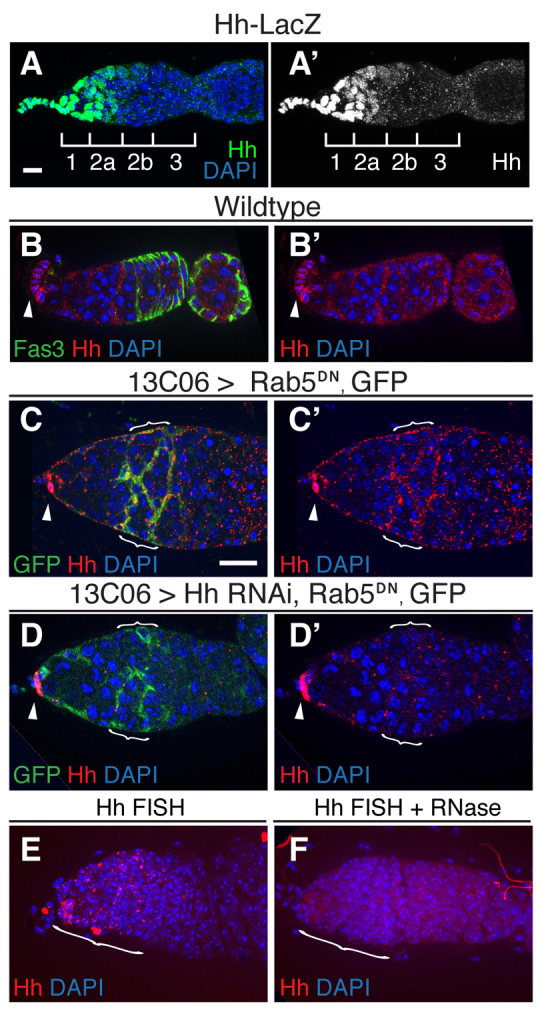
Fig. 4.

Hh is expressed in escort cells. (A,A′) Hh-lacZ expression in the germarium. Tissue is stained for lacZ (β-galactosidase, green) and with DAPI (blue). Regions 1, 2a, 2b and 3 are indicated. (B,B′) Wild-type germarium stained for Fas3 (green) to highlight follicle cell membranes, Hh (red) and with DAPI. Hh signal is bright in cap cells (arrowheads). (C-D′) Germaria in which GFP and Rab5DN are driven in escort cells by 13C06 2 days after flies were shifted to 29°C to repress tub-Gal80ts and promote Gal4 activity. Tissue is stained for GFP (green) to visualize the extent of Gal4 expression, Hh (red) and with DAPI. (C) Hh puncta are abundant on escort cell membranes in region 2a, where Gal4 expression is high (brackets), and sparse in region 1 where Gal4 expression is lower. In addition, as in wild-type germaria, Hh signal is bright in cap cells (arrowheads). Co-expression of HhRNAi with Rab5DN (D) significantly decreases Hh staining in escort cells (brackets) but not cap cells (arrowhead). (E,F) Wild-type germaria stained with a FISH probe for hh transcript (red) and with DAPI. Pretreatment of tissue with RNase (F) eliminates the signal, demonstrating that the FISH probe is specific for an RNA target. Anterior is to the left. Scale bars: 5 μm.
