Fig. 4.
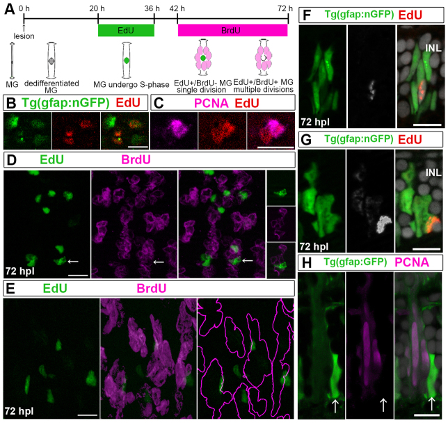
Injury-induced Müller glia divide asymmetrically once to generate neurogenic clusters. (A) Sequential labeling with EdU and BrdU. Two predicted alternative results are depicted: if Müller glia (MG) divide only once, they incorporate only EdU (green); if they undergo multiple divisions, they incorporate both EdU and BrdU (green+magenta=white). (B) Some nGFP+ (green) nuclei in flat-mounted retinas at 42 hpl are EdU+ (red). (C) Pair of EdU+ (red) nuclei at 42 hpl, of which only one is PCNA+ (magenta). (D) Optical z-stack projection at 72 hpl: clusters of BrdU+ nuclei (magenta) with few EdU+ nuclei (green). Right panels: single optical section of EdU+/nGFP+/BrdU- nucleus (arrows). (E) Lateral view of BrdU+ nuclei (magenta) clusters with a single EdU+ (green)/BrdU-nucleus, reconstructed from optical Z-stack. Right panel: BrdU+ clusters (magenta outline). (F) Cluster with single EdU+ cell (white/red) at 72 hpl. (G) Cluster of weakly EdU+ cells (white/red) with one strongly EdU+ cell. (H) Cluster with one PCNA-/strongly GFP+ (green) cell (arrows) and PCNA+ (magenta)/weakly GFP+ progenitors. Scale bars: 10 μm.

