Fig. 7.
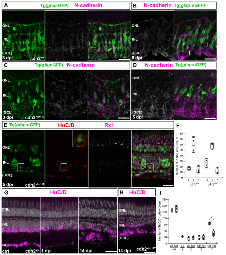
Regeneration of retinal ganglion cells is reduced in cdh2+/m117 heterozygotes. (A,C) At 3 dpi, GFP+/N-cadherin+ (white/magenta) basal Müller glia processes collapse in siblings (A) and hets (C). (B,D) N-cadherin+ (white/magenta) neurogenic clusters in sib retinas (B, arrows) and abnormal clusters in hets (D) at 5 dpi. (E) Regenerated nGFP+/Rx1-/HuC/D+ (red) inner retinal neuron (boxed; enlarged in inset) in het at 5 dpi. (F) Counts of PCNA+ cells in sib and cdh2+/m117. (G) HuC/D+ (magenta) neurons are destroyed at 1 dpi; some regenerate by 14 dpi. (H) Regenerated HuC/D+ neurons in hets. (I) Counts of HuC/D+ neurons. *P<0.0001. Box plots: median, 25th and 75th percentiles; whiskers show maximum and minimum data points. Scale bars: 20 μm.

