
Fig. 6.
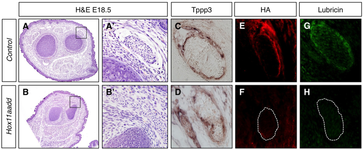
In the absence of Hox11 genes, tendon structure is abnormal and tendon sheath extracellular matrix is not present. (A-H) Transverse sections through the zeugopod of control (A,A′,C,E,G) and Hoxa11/d11 mutant (B,B′,D,F,H) forelimbs at E18.5. Hematoxylin and Eosin (H&E) staining (A-B′) shows abnormal tendon histology in Hox11 mutants. A′ and B′ show higher magnifications of the boxed regions in A and B, respectively. In situ hybridization for the tendon sheath marker Tppp3 (C,D) demonstrates presence of tendon sheath cells. Staining for components of the tendon extracellular matrix hyaluronic acid (HA) (red; E,F) and lubricin (green; G,H) is dramatically reduced surrounding the tendon of Hox11 mutants. In F and H, the tendon analogous to the one shown in E and G is outlined.