Fig. 7.
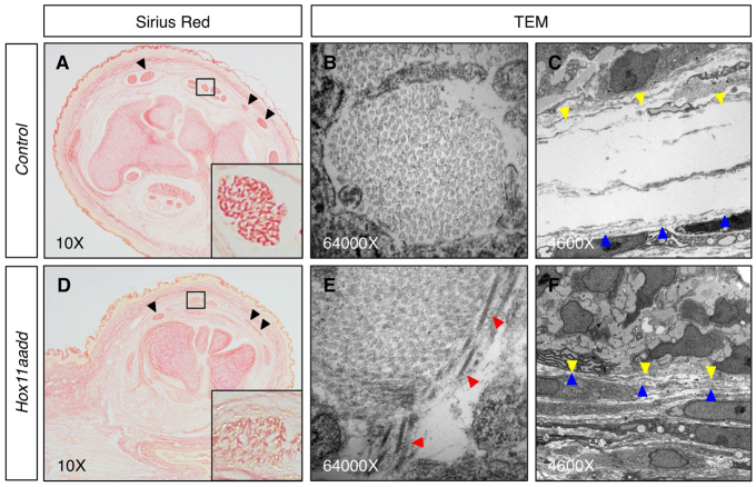
Tendons of Hox11 mutants have disorganized collagen fibers. (A,D) Sirius Red staining for collagen is reduced in Hox11 double-mutant (D) zeugopod tendons at E18.5 compared with control (A). Black arrowheads indicate three dorsal tendons. Insets show higher magnifications of boxed regions. (B,C,E,F) Transmission electron microscopy (TEM) images of collagen fibers in control (B) and Hox11 mutant (E) forelimb zeugopod tendon show disorganization of collagen in Hox11 mutant. Red arrowheads point to collagen fibers running parallel to the plane of section, opposite to the normal orientation. Synovial fluid-filled space surrounding the tendon in control sections (C) is not observed around tendons in Hox11 mutants (F). Yellow arrowheads indicate boundary of tendon fibroblasts. Blue arrowheads indicate boundary of tendon sheath cells.

