Fig. 5.
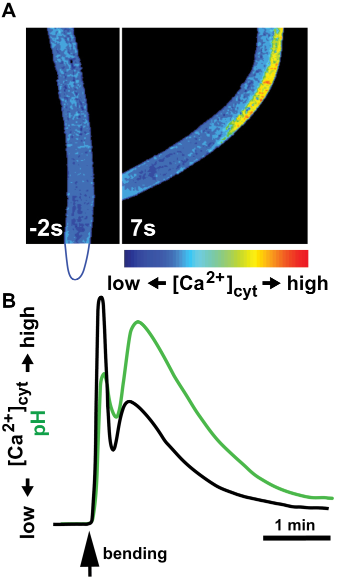
Ion signalling in roots in response to mechanical bending. (A) Arabidopsis root expressing the FRET-based Ca2+ biosensor yellow cameleon 3.6 (Monshausen et al., 2009) is bent to the side with the help of a glass capillary. The position of the root tip (not in the field of view) is outlined in blue below the left panel. Roots exhibit low resting [Ca2+]cyt prior to bending (left) and a rapid increase in [Ca2+]cyt after bending on the stretched (convex) side but not the compressed (concave) side of the roots (right). (B) Kinetics of mechanically triggered [Ca2+]cyt changes in root epidermal cells are echoed by the kinetics of changes in extracellular pH monitored using the fluorescent pH sensor fluorescein conjugated to dextran (based on Monshausen et al., 2009).

