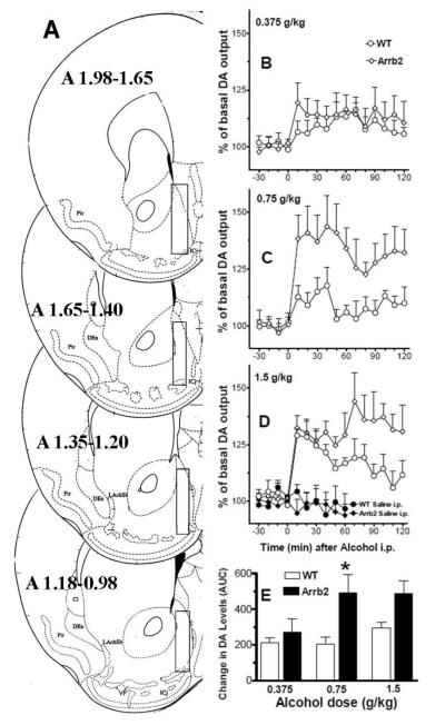
Figure 1. Maximum accumbal DA release is reached at a lower alcohol dose in Arrb2 knockout mice.
Wt and mice lacking Arrb2 were administered alcohol i.p. and DA levels were measured at by in vivo microdialysis in the shell of nucleus accumbens. Panel A shows the brain microdialysis probe placements. Forebrain sections, redrawn from Paxinos and Franklin (2004), show the limits of the positions (boundaries) of the dialyzing portions of the microdialysis probes (superimposed rectangles) within the nucleus accumbens shell. Only the experiments in which the probes were appropriately located inside the nucleus accumbens shell boundaries have been considered and used for the DA microdialysis results shown in the present study. Panels B, C, and D show the time course of the effects of systemic administration of alcohol at doses of 0.375, 0.75, and 1.5 g/kg, respectively, on extracellular levels of DA in dialysates from wt and Arrb2 mice. Panel E shows these responses expressed as area under the curve. Changes in DA levels are presented as percent of baseline, which was established from three consecutive 10-min samples preceding the alcohol challenge, and each data point represents group average ±SEM. Following injection of 0.75 g/kg alcohol, Arrb2 knockout mice display significantly higher DA levels compared to wt mice, p<0.05. Also, the increase in nucleus accumbens shell DA in the Arrb2 group administered with 0.75, and 1.5 g/kg of alcohol, and the increase in DA obtained in the wt group administered the with 1.5 g/kg, were significantly different (p<0.05) from their respective saline treatment groups (see saline treatment on panel D).

