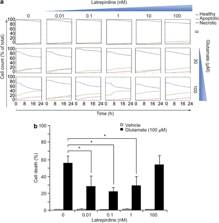
Figure 1.
Latrepirdine pretreatment mediates neuroprotection against excitotoxic injury at (sub)nanomolar concentrations. (a) High-content time-lapse screening of cell death following glutamate excitation. Murine cerebellar granular neurons plated in a 96-well plate were pretreated with a range of concentrations of latrepirdine (0.01–100 nM) for 24 h as indicated. Cells were stained with Hoechst 1 h before treatment with glutamate/glycine (for 10 min at indicated concentrations) after which cells were washed twice with high Mg2+ buffer and preconditioned medium (now containing PI) was replaced. The plate was then immediately placed within the Cellomics imaging chamber (Time 0) and imaged at 1-h intervals over 24 h. Cells were categorized and analysis was carried out using Cell Profiler as described in the Materials and Methods. Data presented are representative traces from thousand of cells, and experiments were carried out on three independent neuronal cultures. (b) Murine cerebellar granular neurons were plated in 24-well plates and following pretreatment with latrepirdine (0.01–100 nM as indicated) for 24 h, cells were exposed to glutamate/glycine 100 μM/10 μM for 10 min. After treatment, cells were washed twice with high Mg2+ buffer and incubated in preconditioned medium for a further 24 h. Pyknotic nuclei were counted as apoptotic, as determined by Hoechst 33358 staining (1 μg ml−1) and expressed as a percentage of total (n=4 independent experiments in triplicate). Data are presented as mean±s.e.m. *P⩽0.001 indicates difference between glutamate-only treated and latrepirdine (0.01–1 nM)-pretreated glutamate-treated neurons.

