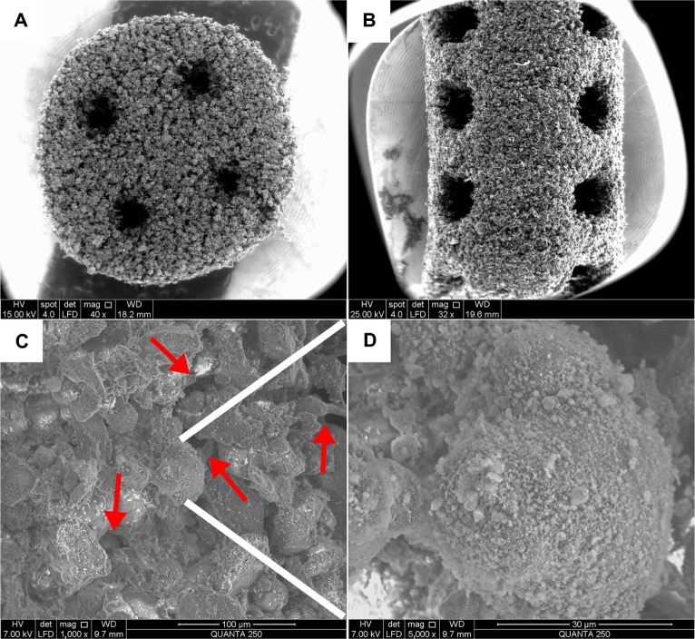
Figure 2.
Scanning electronic microscopy micrographs of the scaffolds.
Notes: (A) Base (×40) and (B) peripheral (×32) areas of the nano-hydroxyapatite/poly-ε-caprolactone (PCL) scaffolds with well-ordered and interconnected macropores from 600 μm to 800 μm. (C) Microstructure of the scaffolds (×1,000). The micropores (red arrows) appeared among PCL particles with pore sizes of several microns in diameter. (D) The PCL particles were evenly attached to the nanoscale hydroxyapatite particles (×5,000).

