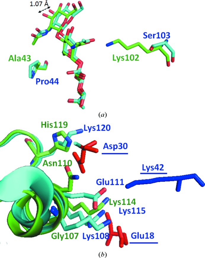
Figure 4.
(a) Interaction of (GlcNAc)3 with binding sites. (GlcNAc)3 in MLL and VPL is shown in green and cyan, respectively. (b) Comparison of the amino acids that contribute to dimer formation. Amino acids in MLL and VPL are shown in green and cyan, respectively. The amino acids derived from the second subunit of the VPL dimer are underlined.

