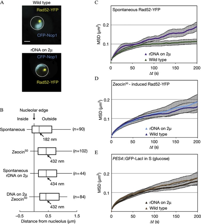
Figure 4.
Spontaneous Rad52-YFP foci are constrained by the nucleolus. (A) Representative images of wild-type cells (GA5820) and those harbouring the rDNA on a multicopy plasmid (GA7997), both expressing CFP-Nop1. The CFP-Nop1 signal in the strain carrying the rDNA on a plasmid was ∼10-fold lower than in wild type, and thus brightness is adjusted. Scale bar, 1 μm. (B) Distances between Rad52-YFP foci and the nucleolus marked by CFP-Nop1 in GA5820 and GA7997. The number of nuclei analysed (n) is given for each condition. (C,D) MSD analyses of spontaneous (C) and Zeocin-induced (D) Rad52-YFP foci in both wild-type cells with (GA5820; 21 and 28 movies, respectively) or without the rDNA on a 2 μ plasmid (GA7997; 16 and 26 movies, respectively). 50 μg/ml Zeocin treatment was for 1 h. (E) MSD analysis of the LacO-tagged PES4 locus in wild type (GA1461; 20 movies) and in cells with the 5S and 35S on a 2 μ plasmid (GA8147; 22 movies). Analyses were done on S-phase cells. The error bars on the MSD curves represent the s.e. CFP, cyan fluorescent protein; MSD, mean square displacement; rDNA, ribosomal DNA; YFP, yellow fluorescent protein.

