
Fig. 5.
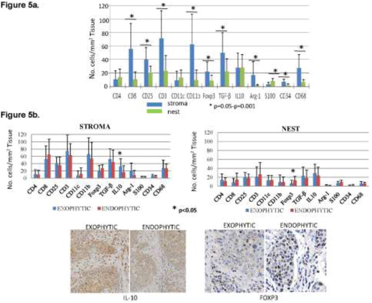
Inflammatory cells in the tumor stroma vs. tumor nests. (a) Numbers of immune cells positive for various surface markers in the tumor stroma and tumor nests (n=30). Asterisks indicate significant differences at p<0.05 to p<0.001. (b) Absolute numbers of immune cells positively stained for different markers in the stroma and nests in tumors with exophytic (n=22) and endophytic (n=8) tumors. On the right, representative tumor sections showing the presence of IL-10+ and FOXP3+ cells in the tumor stroma or tumor nests. Mag ×200 for IL-10+ and ×400 for FOXP3 staining.