
Fig. 7.
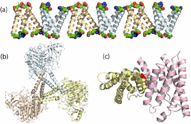
Bilayer and non-bilayer lattices in membrane protein crystals. a) Despite the bilayer lattice for the Influenza A M2 protein (3BKD) significant electrostatic interactions (space-filling Arg and Glu residues) between antiparallel tetramers appear to distort the tetramer helices (Stouffer et al. 2008). b) The non-bilayer lattice of estrone sulfatase (1P49) showing three monomers with nearly orthogonal TM helices and significant inter-monomer interactions (Hernandez-Guzman et al. 2003). c) The energy coupling factor-type riboflavin transporter (3P5N) also forms a lattice in which the TM domain of one protein is rotated by ~90° with respect to its neighbor and inter-protein interactions include helices that pack together via an Ala motif (Zhang et al. 2010).