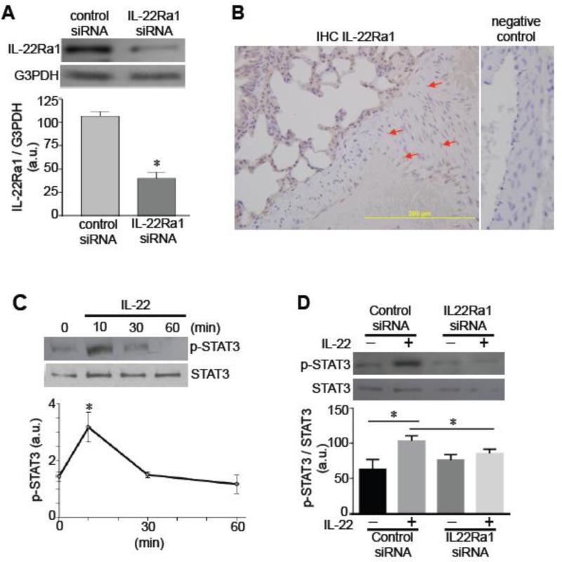
Fig. 1. Expression of functional IL-22 receptor in pulmonary artery SMCs.
[A] Human pulmonary artery SMCs were transfected with control siRNA or IL-22Ra1 siRNA. Cell lysates were subjected to immunoblotting with the IL-22Ra1 antibody. The glyceraldehyde-3-phosphate dehydrogenase (G3PDH) expression was monitored as a control. The bar graph represents means ± SEM (n = 6) of the ratio of IL-22Ra1 to G3PDH as expressed in arbitrary units (a.u.). The symbol * denotes values significantly different from control at P<0.05. [B] Rat lungs were immersed in buffered 10% paraformaldehyde, and embedded in paraffin for IHC with the IL-22Ra1 antibody. Brown stains in the pulmonary artery medial layer are indicated by the red arrows. [C] Human pulmonary artery SMCs were treated with IL-22 (10 ng/ml) for durations indicated. Cell lysates were prepared and subjected to immunoblotting for phosphorylated STAT3 (p-STAT3). The line graph represents means ± SEM (n = 3). * denotes values significantly different from control at P<0.05. [D] Human pulmonary artery SMCs were transfected with control siRNA or IL-22Ra1 siRNA, then treated with IL-22 for 10 min to monitor phosphorylated STAT3 by immunoblotting. The bar graph represents means ± SEM (n = 4). * denotes values significantly different from each other at P<0.05.

