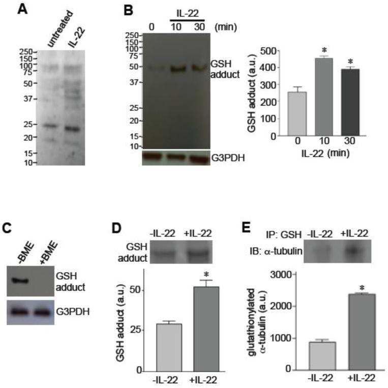
Fig. 4. IL-22 promotes protein glutathionylation.
[A] Human pulmonary artery SMCS were treated with IL-22 (10 ng/ml) for 10 min, and the levels of GSH-protein adducts were monitored by non-reducing SDS-PAGE and immunoblotting using a GSH antibody from Millipore. [B] Human pulmonary artery SMCS were treated with IL-22 (10 ng/ml), and the levels of GSH-protein adducts were monitored by non-reducing SDS-PAGE and immunoblotting using a GSH antibody from Santa Cruz Biotechnology. [C] Immunoblotting for detecting GSH-protein adducts with the GSH antibody from Santa Cruz was performed without or with BME in SDS-PAGE. [D] Lysates from cells treated without or with IL-22 for 10 min were immunoprecipitated with the GSH antibody from Santa Cruz, followed by non-reducing SDS-PAGE and Coomassie blue staining. [E] Lysates from cells treated without or with IL-22 for 10 min were immunoprecipitated (IP) with the GSH antibody from Santa Cruz, followed by non-reducing SDS-PAGE and immunoblotting (IB) with the α-tubulin antibody to monitor the levels of glutathionylated α-tubulin. * denotes values significantly different from untreated control at P<0.05 (n = 3).

