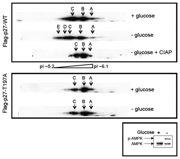
Figure 3. Activation of AMPK signaling increases p27 phosphorylation at T197.
NIH3T3 cells were transfected with Flag-p27-WT or Flag-p27-T197A constructs, grown in the presence or absence of glucose, and then utilized to generate whole-cell lysates that were untreated or treated with CIAP. Lysates were then separated on 2-D gels and immunoblotted for p27 or separated by SDS-PAGE and immunoblotted for AMPK-α and phospho-AMPK-α (T172). Arrows A, B, and C indicate p27 isoforms detectable under all conditions, and arrows D and E indicate additional p27 isoforms with lower pIs (indicative of phosphorylation) that are detectable only upon glucose deprivation, which activates AMPK signaling.

