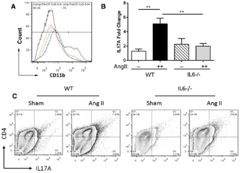
Figure 2. IL-6 deficiency reduced Ang II-induced macrophage and Th17 recruitment.

Age matched WT and IL-6−/− mice were treated with Ang II or saline (sham) for 14 d. (A) Flow cytometric analysis of CD11b-positive macrophages was performed using disassociated aortic cells and the number of CD11b-positive cells was measured. Black curve: Sham-treated WT. Blue curve: Ang II-treated WT. Red curve: Sham-treated IL-6−/−. Green curve: Ang II-treated IL-6−/−. n=4 in each group. (B) IL-17A expression was analyzed using Q-RT-PCR. White bar: Sham-treated WT. Black bar: Ang II-treated WT. Cross bars: Sham-treated IL-6−/−. Grey bar: Ang II-treated IL-6−/−. n= 3–5 in each group. *, p<0.05.**, p<0.01.(C) Flow cytometric analysis of CD4-positive and IL-17A-positive cells was performed and number of double-positive cells was measured. Representative panels corresponding to each group are shown (n=6). IL-6−/− showed abated Th17 recruitment to the aorta.
