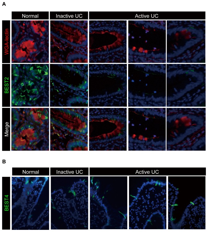
Figure 5. Expression of BEST2 is markedly down-regulated in active lesions of UC.
The immunohistochemical analysis of BEST2 and BEST4 expression using colonic tissues of UC patients is shown. Representative data obtained from three subjects in each group are presented. (A) Expression of BEST2 is down-regulated in the active lesions of UC patients, where goblet cell mucins are depleted. The double staining of BEST2 (green) and WGA-lectin (red) using tissues from the normal human colon or from active and inactive lesions of UC patients is shown (Original magnification 400x). The red staining of WGA-lectin represents goblet cell mucins. Each series represents staining results obtained from a different patient (Original magnification 400x). (B) The expression of BEST4 is maintained at the surface epithelium of active, as well as inactive, lesions in UC patients. Single staining of BEST4 (green) using tissues from the normal human colon or from active and inactive lesions of UC patients is shown (Original magnification 400x). Each figure represents a staining result obtained from a different patient.

