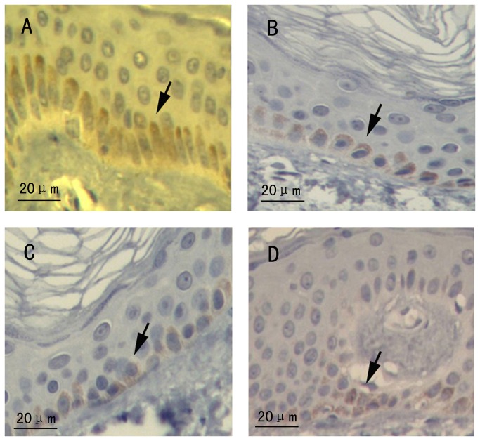
Figure 4. Immunohistochemistry staining of ABCB6 observed under light microscopy.
(A) Section of skin tissue from the normal control. (B) Section of hyperpigmented tissue from the proband with c. 1663 C>A. (C) Section of hypopigmented area from the proband. (D) Section of skin tissue from the patient with c.459 delC. (A-D) All of the melanocytes were positive immunoreactivity for ABCB6. Arrows indicate melanocytes.

