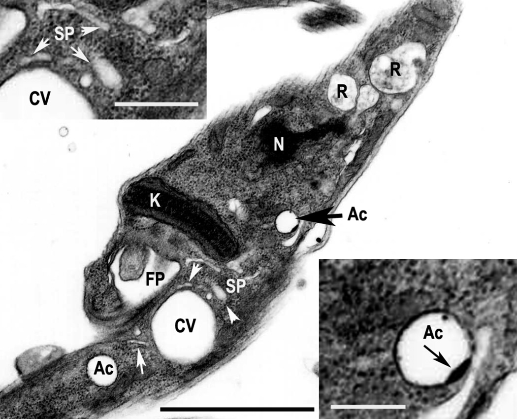
Figure 1. Acidocalcisomes of T. cruzi.
Epimastigotes were observed by transmission electron microscopy. Notations are flagellar pocket (FP), acidocalcisome (Ac), contractile vacuole (CV), spongiome (SP), nucleus (N) and reservosome (R). Insets show the spongiome (left) and one acidocalcisome (right) at higher magnification. Note the electron-dense inclusion (black arrow) in the membrane of the acidocalcisome, which also has an electron dense periphery. White arrows show the tubules of the spongiome. Bar = 2.5 µm (main picture), and 0.2 µm (inset).

