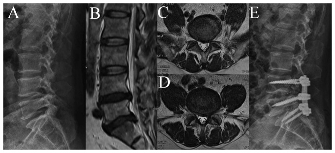
Figure 1. A 54-years-old man, diagnosed as LDH and left foot drop.

(A) Preoperative radiography. (B) Preoperative mid-sagittal MRI showed LDH on L4-S1. (C) Preoperative axial MRI of L4-5 showed the left L5 nerve root compression. (D) Preoperative axial MRI of L5-S1 showed the left S1 nerve root compression. (E) Postoperative radiography.
