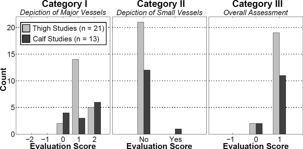
Figure 6.
Histograms of the results of the radiological evaluation for the thigh and calf studies. Categories I – III are defined in Table 2. In Categories I and III, the null hypothesis of “no improvement” was rejected in a statistically significant way, showing that images reconstructed with vascular masking have a radiological improvement compared to images reconstructed with conventional masking. In Category II, there was no significant loss of vessels due to masking.

