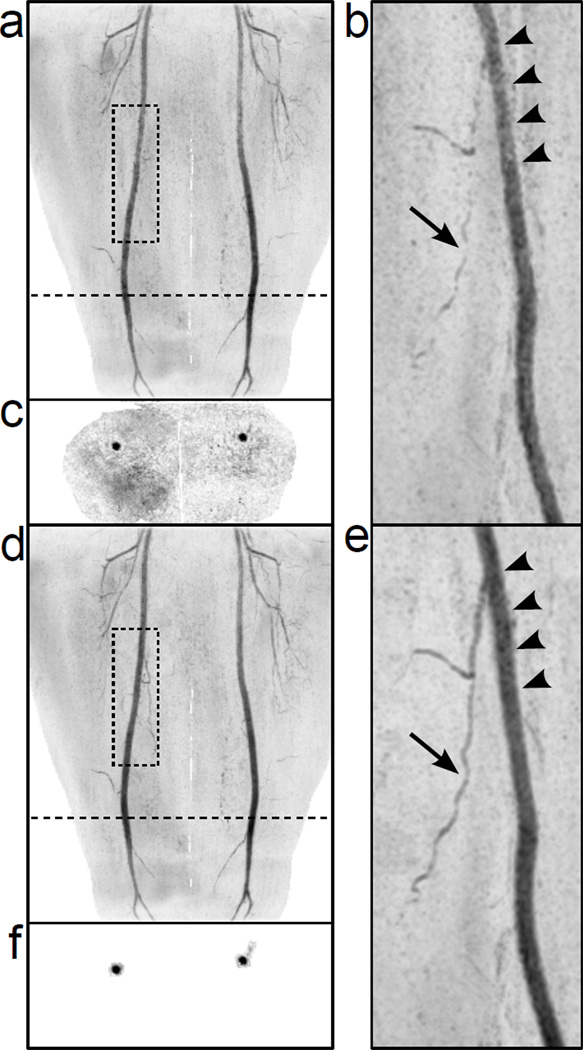
Figure 7.
Full-FOV and rotated targeted MIPs of R = 8 2D SENSE-accelerated 3D CE-MRA thigh angiograms reconstructed with the conventional masking technique (a-b) and the new vascular masking technique (d-e). Representative axial slices of the datasets are shown in (c) and (f). The position of the axial slices is shown by the dotted line on the full-FOV MIPs. The image reconstructed with the vascular mask (e) shows improved luminal signal smoothness (arrow heads) and better small vessel conspicuity (arrow) vs. conventional masking (b).

