
Figure 2. Placental and embryonic mRNA expression levels of genes involved in β-carotene cleavage and retinoid homeostasis following β-carotene supplementation at 13.5 dpc.
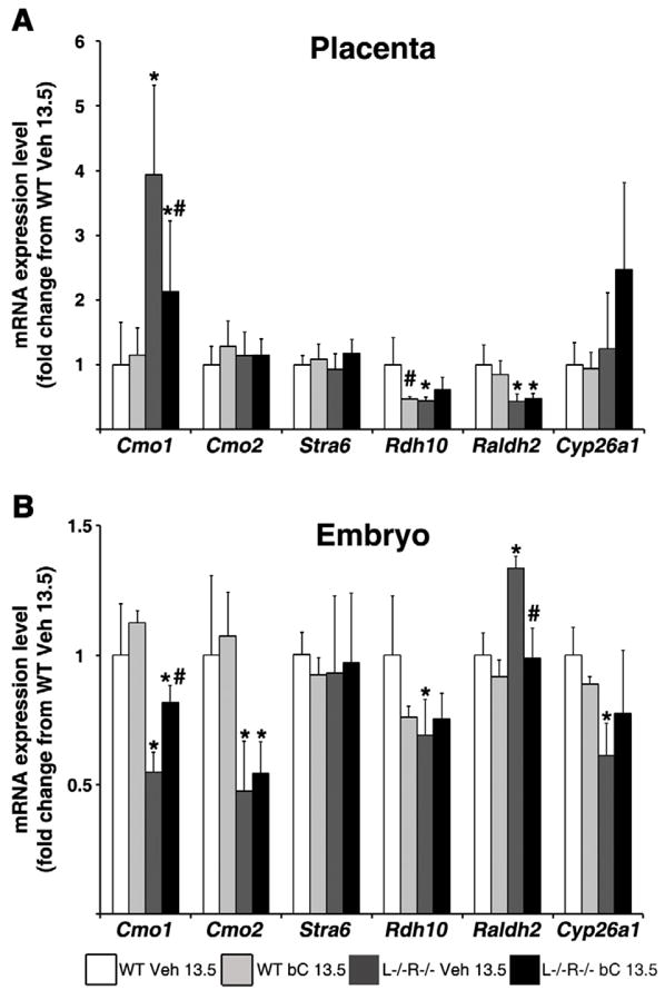
qRT-PCR analysis was performed using mRNA from 14.5 dpc placentas (A) and embryos (B) from wild-type (WT) and Lrat−/−Rbp−/− (L−/−R−/−) dams treated at 13.5 dpc with Vehicle (Veh 13.5) or β-carotene (bC 13.5). Tissues of WT Veh 13.5 were set as calibrator at 1. Data are presented as mean ± SD fold of WT Veh 13.5. Sample size, n=5–10 placentas or embryos/group (from 3–5 dams/group). Statistical analysis was performed by two-way ANOVA with genotype and treatment as factors, followed by LSD or Tamhane’s post hoc analysis. Individual comparisons then were made by Student’s t test. *, p<0.05 vs. WT; #, p<0.05 vs. Veh.