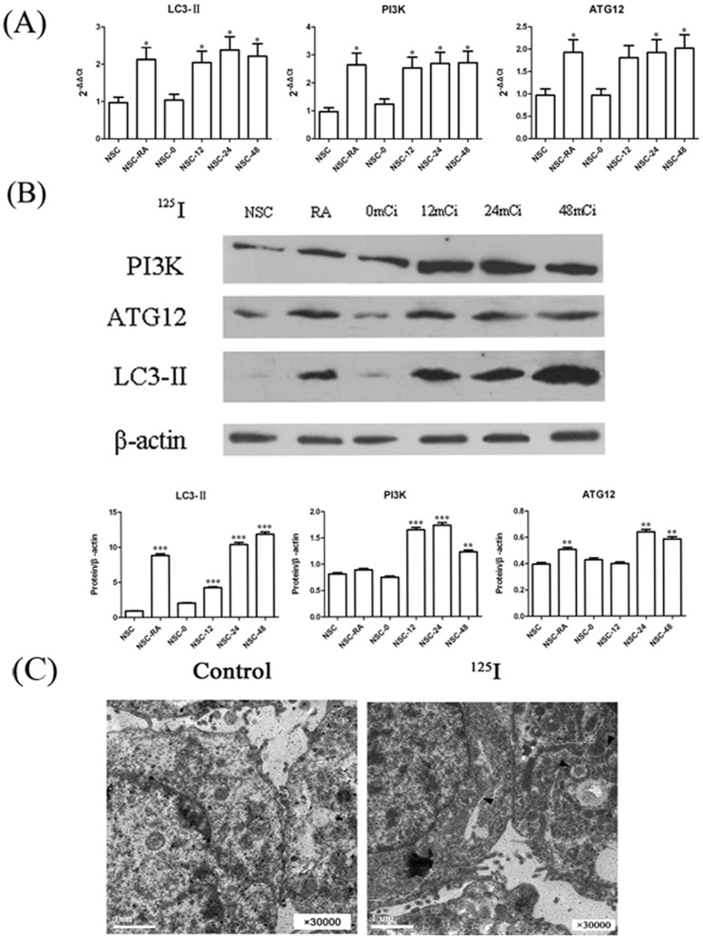
Figure 2. Induction of autophagy related proteins in neurons.
(A) Dose response assay. Neural cells were treated with a range of 125I radiation (12mCi ∼48mCi) for 5 days. Relative mRNA expressions were measured by qPCR. The data were shown as mean value ± SEM., and they were representative of three independent experiments. (B) Neural cells were treated with a range of 125I radiation (12mCi ∼48mCi) for 5 days. Levels of proteins expression were measured by immunoblot ananlysis using antibodies against LC3II, Atg12, PI3K and beta-actin. Neuron cells treated with RAPA were as positive controls and untreated neurons were as negative controls. Results here were representative of three independent experiments. (C) Electron microscopic features of rat neural cells treated with 125I for 24 h. Arrows indicated autophagosomes.

