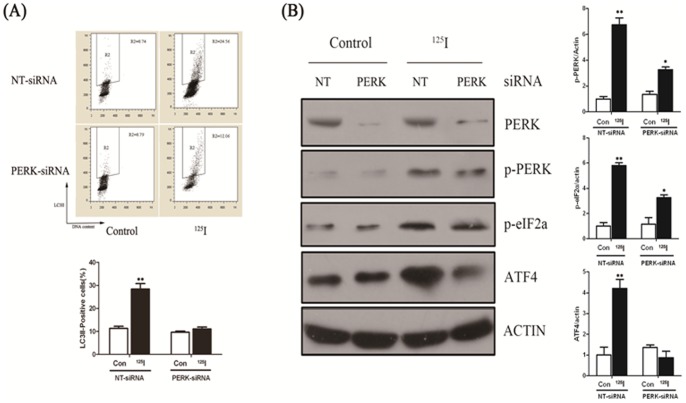
Figure 4. Neuron cells were transfected with RNAi against PERK and control (NT).
(A) 16 h later, cells were allowed to recover for 3 days, and LC3II/DNA flowcytometry analyses after indicated period of time. The gate (R2) represented LC3II positive cells in each figure. * indicated statistically significant differences (p<0.05) between the levels of 125I treated and without treatment (NT) in cells transfected with PERK-siRNA or NT-siRNA. (B) Protein levels of PERK, p-PERK, p-eIF2α, ATF4 and β-actin were measured by immunoblot analysis in nueral cells transfected with RNAi specific to PERK and control cells (NT) which were radiated with 125I (24mCi) for 5 days. The expressions of p-PERK, p-eIF2α and ATF4 significantly decreased in response to 125I irradiation in neurons transfected with siRNA against PERK. Results were representative of three independent experiments. (B) Quantification of the mentioned protein levels in cells transfected with RNAi against PERK and control (NT) following 125I radiation. *, p<0.05, **, p<0.01, the error bar represented the SEM.

