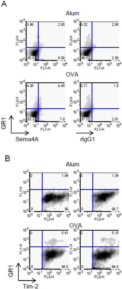
Figure 4.
Subsets of lung GR1+ cells express Sema4A and Tim-2. Single cell suspensions obtained from lungs of Alum- or OVA-treated mice were analyzed for GR1, Sema4A, and Tim-2 expression using corresponding Abs defined in Materials and Methods section. (A) Approximately a half of lung GR1+ cells co-expressed Sema4A. (B) Tim-2+ cells were selected for further analysis based on SSC-Fl3 dot plots (not shown) and then re-gated on Fl1-Fl3 density plots. Note that GR1+Tim2+ cells appear only under inflammatory lung conditions.

