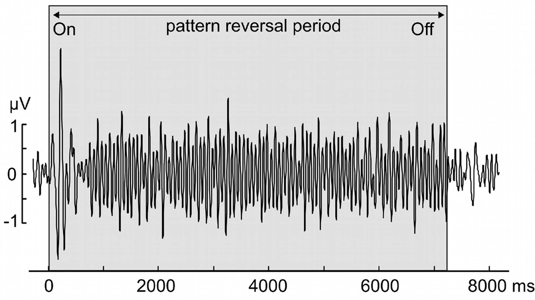
Figure 2.
Example of the time-domain representation of the ssVEP recorded at the occipital midline electrode location (Oz) when viewing the luminance stimulus, from one representative participant, in the 14 Hz condition. The signal is averaged across experimental phases and conditions, and shows the entrainment of visual cortical areas at the reversal rate of 14 cycles per second.

