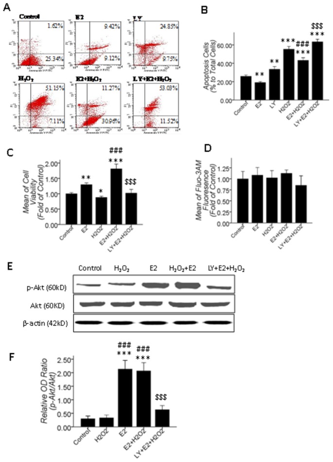
Figure 6. 10 μM βE2 pretreatment for 0.5 hrs protected primary cultured SD rat retinal cells from apoptosis induced by 100 μM H2O2 treatment for 24 hrs.

The PI3K/Akt pathway mediated this process, but the alteration in [Ca2+]i was undetectable. A: The Annexin V/Propidium Iodide staining apoptosis assay; B: Quantitative data of A; C and D: Cell viability and [Ca2+]i quantitative data; 10 μM βE2 pretreatment for 0.5 hrs significantly restored the decrease in cell viability and apoptosis, which was significantly inhibited by 10 μM LY (B, C), but the [Ca2+]i was not significantly altered in all treated groups (D); E: Western blot results, 10 μM βE2 pretreatment for 0.5 hrs promoted p-Akt level, which was inhibited by 10 μM LY pretreatment for 0.5 hrs before βE2 and H2O2 co-treatment. F: Quantitative data of E. Values shown are the Mean ±SD. *represents P<0.05, **represents P<0.01 and ***represents P<0.001 compared with the control group by the T-test or one-way ANOVA statistical analysis; ### represents P<0.001 compared with the H2O2 application group by one-way ANOVA statistical analysis; $$$ represents P<0.001 compared with the βE2 and H2O2 co-application group by one-way ANOVA statistical analysis. (B, C, D: n indicates 3 independent replicates with 4 samples per condition per experiment; F: n indicates 3 independent replicates.).
