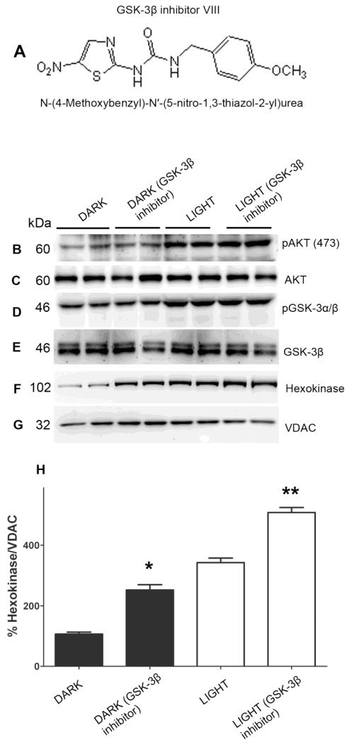
Figure 6. GSK-3β regulation of binding of HK-II to mitochondria.
Chemical structure of GSK-3β inhibitor N-(4-Methoxybenzyl)-N’-(5-nitro-1,3-thiazol-2-yl)urea (A). Retinal explants from dark-adapted rats were incubated with and without GSK-3β inhibitor. The ex vivo explants were exposed to light or kept in the dark for 30 min. Mitochondria were prepared and the proteins were immunoblotted with anti-pAkt (B), anti-Akt (C), anti-pGSK-3α/β (D), anti-GSK3β (E), anti-HK-II (F), and anti-VDAC (G) antibodies. Densitometric analysis of HK-II normalized to VDAC (H). The dark control was set as 100 percent. Values are mean + SD, n=2. *p<0.01 compared to dark-adapted control. **p<0.001 compared to light-adapted control. Mitochondria were prepared from two independent sets of light- and dark-adapted retinas. Each set had at least 20 rats (10 animals for light- and 10 animals for dark-adaptation).

