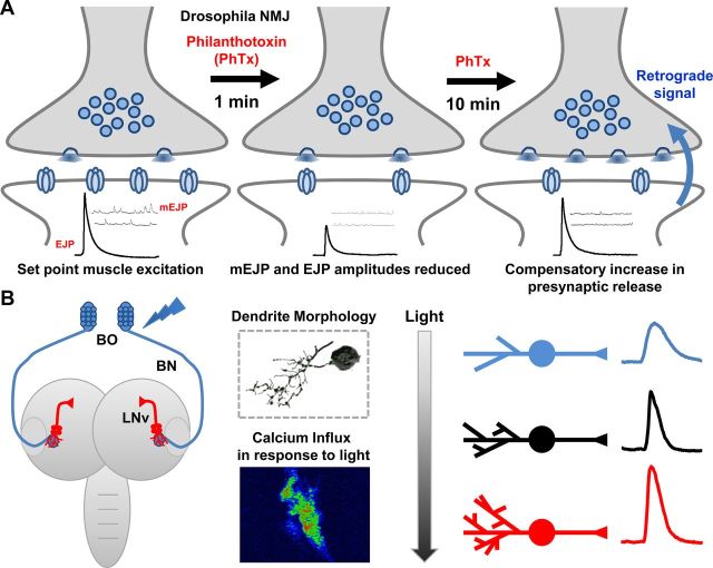
Figure 2.
Drosophila models of homeostatic synaptic plasticity. A, Schematic of homeostatic compensation after application of the postsynaptic glutamate receptor antagonist philanthotoxin (PhTx) to the larval NMJ. Application of PhTx initially causes an ∼50% decrease in miniature excitatory junction potential (mEJP) amplitude and a parallel ∼50% decrease in evoked excitatory junction potential (EJP) amplitude. After 10 min incubation in PhTx, mEJP amplitudes remain depressed whereas EJP amplitudes increase to baseline values resulting from a homeostatic enhancement of presynaptic neurotransmitter release (quantal content). B, Schematic of the larval visual circuit; the light sensing BO sends an axon projection (BN) into the larval brain where it synapses with the LNv (left). LNvs exhibit activity-dependent structural and functional plasticity, demonstrated by 3D tracing and calcium imaging (middle). Light exposure bidirectionally alters dendrite length and synaptic activity (right).

