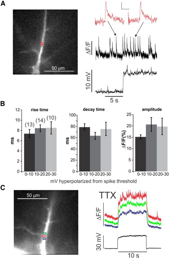
Figure 1.

Changing membrane potential increases the frequency of spontaneous sparks but does not change their kinetic parameters or change them into propagating events. A, Spontaneous events detected at a dendritic location (red ROI). The fluorescence image shows a pyramidal neuron filled with OGB-1. During the 10 s trial, the membrane potential was depolarized by 10 mV, increasing the rate of events. The inset shows two events, one before and one after the depolarization. There was no change in the kinetics or amplitude. Calibration: inset, ΔF/F, 200 ms. B, Histograms of rise times (10–90%), half-decay times, and amplitudes of spontaneous events measured at different potentials below the threshold for spike generation. There were no significant changes (p > 0.3 in all cases). Numbers in parentheses above bars are numbers of measured events, same for all three histograms. C, [Ca2+]i changes measured at three neighboring locations in the dendrites in response to a 30 mV step depolarization in ACSF containing 1 μm TTX. The rate of events increased at the center location (red trace) but did not change at the neighboring sites. The steady increase in [Ca2+]i at all three sites was attributable to voltage-dependent Ca2+ entry.
