
Figure 9.
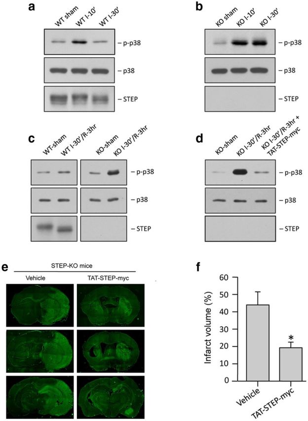
Administration of TAT-STEP peptide at the onset of reperfusion reduces p38 MAPK phosphorylation and ischemic brain damage in STEP KO mice. a, Immunoblot analysis of striatal lysates from the ipsilateral side of WT mice, after 10 or 30 min MCAO (n = 3/group). b, Immunoblot analysis of striatal lysates from the ipsilateral side of STEP KO mice, after 10 or 30 min MCAO (n = 3/group). c, Immunoblot analysis of striatal lysates from the ipsilateral side of WT mice and STEP KO mice, after 30 min MCAO and 3 h reperfusion (n = 3/group). d, Immunoblot analysis of striatal lysates from the ipsilateral side of STEP KO mice subjected to 30 min MCAO followed by administration of TAT-STEP-myc peptide (3 nmol/g) and reperfusion for 3 h (n = 3/group). a–d, Blots were probed with anti-phospho-p38 MAPK (top) and reprobed with anti-p38 MAPK (bottom) or anti-STEP antibody (bottom). e, STEP KO mice were subjected to 30 min MCAO, and the peptide was administered at the onset of reperfusion. Representative photomicrographs of coronal brain sections, 24 h after the onset of ischemia, stained with Fluoro-Jade C. f, Total infarct volume was 43.9 ± 7.07% for STEP KO mice compared with 19.3 ± 2.87% in STEP KO mice. *p < 0.05. n = 5.