
Figure 2.
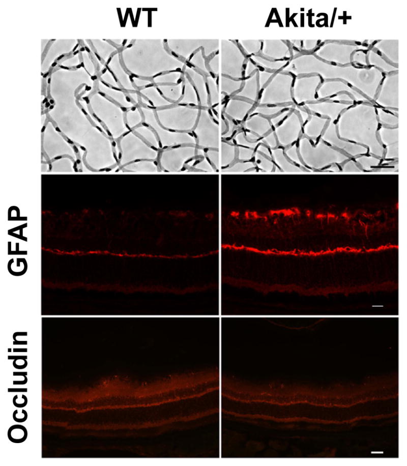
Decreased numbers of pericytes in Akita/+ male mice. Retinas from 7 month old wild-type and Akita/+ mice were prepared by trypsin digest as described in Materials and Methods (scale bar= 20 μM) [11,41]. GFAP and occludin staining of retinal sections from 9 month wild-type and Akita/+ male mice are also shown (scale bar= 50 μM). Note that GFAP expression increased in sections from Akita/+ male mice. Experiments were repeated with eyes from 5 mice with similar results.