
FIGURE 3:
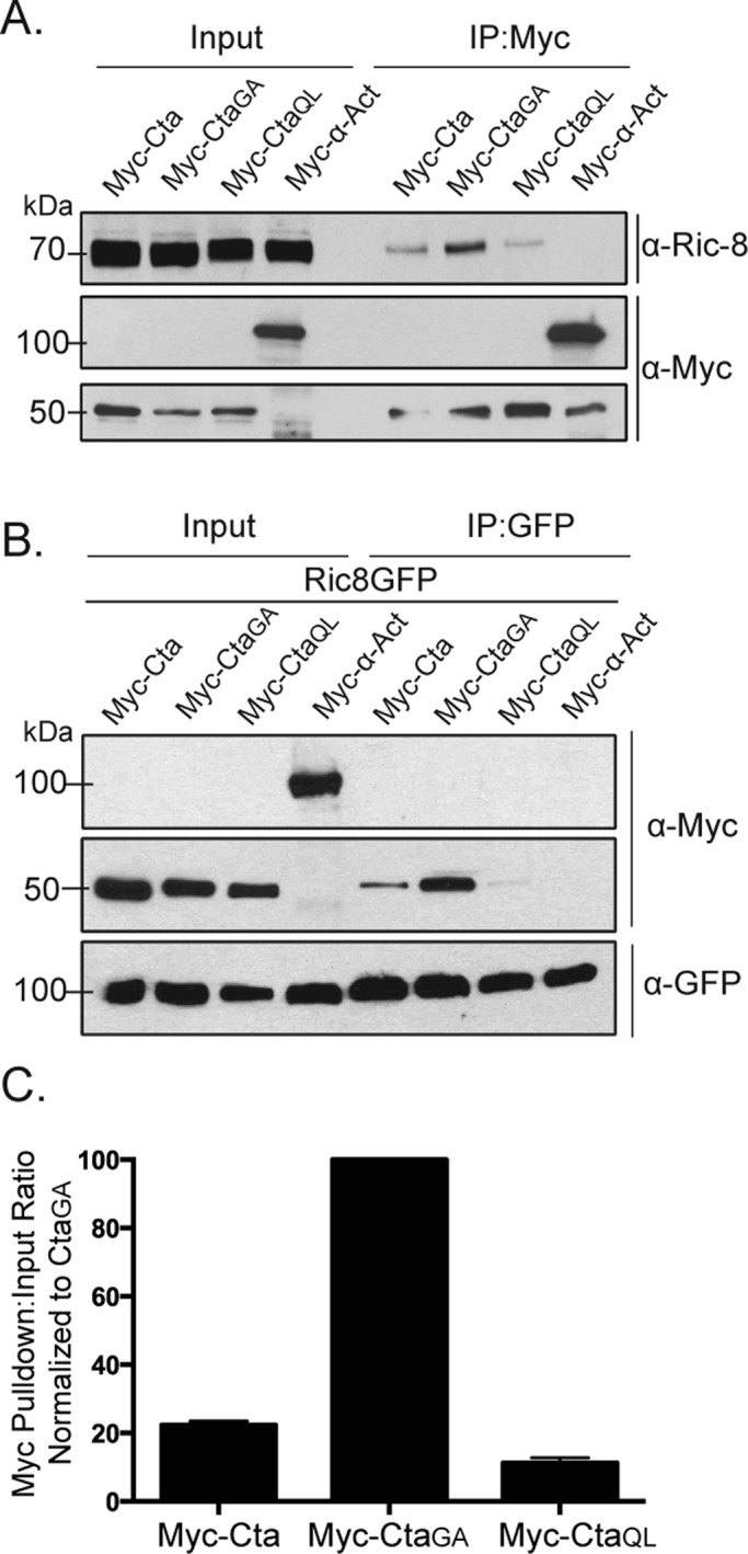
Ric-8 physically interacts with Cta and exhibits higher binding affinity for constitutively inactive Cta. (A) S2 cells were transfected with Myc-Cta, Myc-CtaGA, Myc-CtaQL, or α-actinin (α-Act) as a negative control. IPs were performed using an anti-Myc antibody, and samples were probed with anti–Ric-8 and anti-Myc. (B) S2 cells were transfected with Ric-8–GFP and Myc-Cta, Myc-CtaGA, Myc-CtaQL, or α-Act. IPs were performed with GFP-binding protein and probed with anti-GFP and anti-Myc. (C) Quantification of IPs performed in B. Pull-down:input ratios were determined using quantitative densitometry and normalized against CtaGA (±SEM).