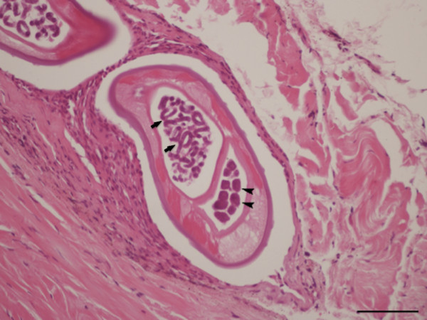
Figure 5.
Periocular tissue, histopathology. In the subconjunctival sac there is a section of a coiled female of Onchocerca lupi, note the uteri filled with eggs containing well-developed microfilariae (arrows) or eggs in earlier stage of development (arrowheads), a faint intestine, atrophied muscle and lateral hypodermal chords (H&E, scale-bar = 100 μm).

