
Figure 1.
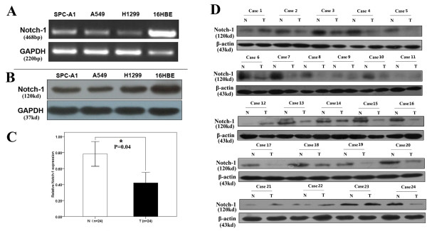
Expression of Notch-1 in LAD Cell lines and tissues. Semi-quantitative Reverse transcription-polymerase chain reaction (A) and Western Blot (B) were used to detect expression of Notch-1 in different cells of lung adenocarcinoma. Brochial epithelial cell was used as control. Weaker expression of Notch-1 was observed in tumor cells. Then, Notch-1 Protein in 24 tissues from surgery which diagnosed as lung adenocarcinoma were detected by Western Blot (C and D). Each adjacent tissue from the same patient was used as control. Most of weaker performance was observed in tumor ones (P = 0.04).