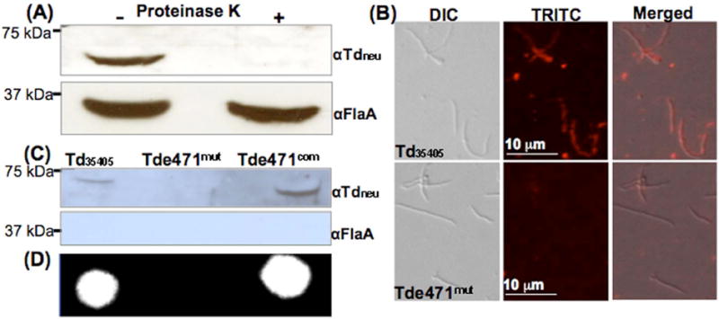
Figure 3. Localization of Tdneu.

(A) Immunoblotting analysis of proteinase K-treated Td35405 whole-cell lysates. The spirochetes were either incubated with proteinase K (240 μg/ml) or PBS at 37°C for 1 h. The resultant samples were separated on SDS-PAGE and then probed with αTdneu and αFlaA. (B) IFA analysis. Td35405 and Tde471mut cells were fixed with methanol, stained with αTdneu, and counterstained with a goat-anti-rat Texas red antibody as previously described. The micrographs were taken under DIC light microcopy or fluorescence microscopy with a tetramethylrhodamine isothiocyanate (TRITC) emission filter, and the resultant images were merged. (C) Detection of Tdneu in the supernatants prepared from the cultures of Td35405, Tde471mut, and Tde471com strains by immunoblotting analysis. FlaA was used as a sample preparation control. (D) Detecting neuraminidase activity in the supernatants by the filter paper spot test as described in Figure 1A.
