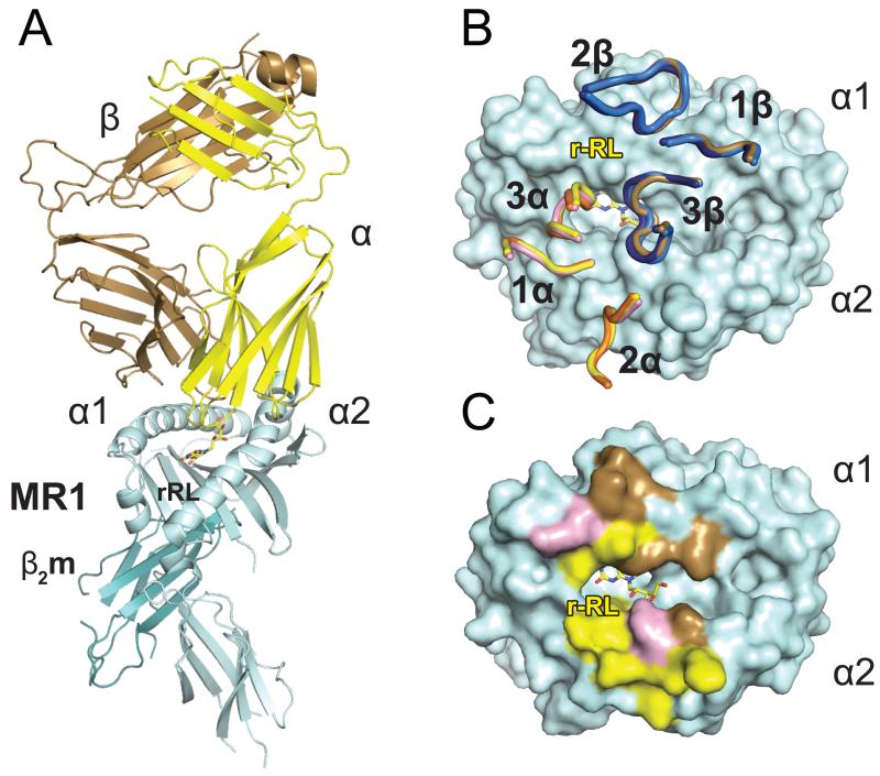
Figure 3.
MAIT TCR recognition of MR1-antigen. A) Cartoon diagram of the ternary complex structure human MAIT TCR/hbMR1 and the MAIT cell stimulatory ligand rRL-6-CH2OH. The TCR α and β chains are shown respectively in yellow and brown colors, MR1 in cyan and β2m in teal. rRL-6-CH2OH is represented as yellow sticks. B) Superposition of the F7 MAIT TCR CDR loops in the complexes with bovine (shown in pink and marine for the α and β chains, respectively) and hbMR1 (shown in yellow and brown for the α and β chains, respectively) and comparison with the loop positioning in the human MAIT/MR1/RL-6-Me-7-OH complex (16) (loops shown in orange and dark blue for the α and β chains, respectively). All three complexes are aligned via MR1 and the respective CDR loops are displayed on top of the hbMR1 surface. C) Footprint of the F7 human MAIT TCR on hbMR1 surface. hbMR1 residues contacting the TCR α chain are shown in yellow and those contacting the β chain are shown in brown. Residues making contacts with both chains are in pink.

