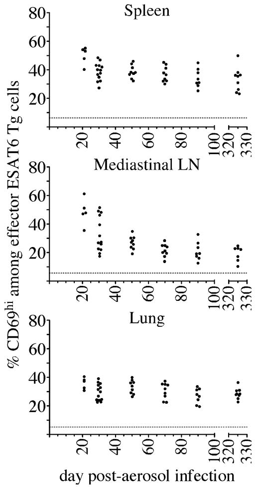
Figure 4. Effector T cells detect similar levels of antigen during Mtb infection.

In vitro differentiated ESAT6 Th1 effectors (1×10⁶) were injected into congenic recipient mice, and mice were analyzed 12 hours after cell transfer on each of the days indicated. The percent of cells that expressed of CD69 on donor ESAT6 Th1 effector T cells in the spleen, MLN, and lung is shown. The datum represents individual mice; the dashed line indicates the mean CD69 expression detected on effector Th1 cells that were transferred into uninfected mice. The data shown were accumulated from three experiments.
