Figure 1.
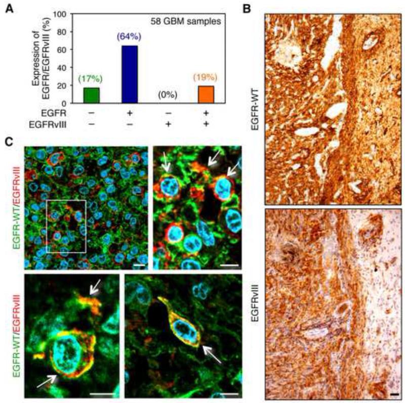
Detection of EGFR and EGFRvIII in primary human glioblastoma. (A) Graphical analysis of immunohistochemical data from Table S1. (B-C) Immunohistochemical staining of a primary human GBM with EGFR-specific (top panel) or EGFRvIII-specific antibody (bottom panel) on consecutive sections (brown, diaminobenzidine; light blue, nuclear counterstain with hemalum). Black scale bar corresponds to 50 μm. (C) Immunofluorescence double-staining of primary GBM tissue sections using EGFR-specific and EGFRvIII-specific antibodies. Cells double-positive for both EGFR-WT and EGFRvIII are indicated by arrows. An enlarged image of region marked with a white square (upper left panel in (C)) is shown on the right side. Lower panels demonstrate EGFR-WT/EGFRvIII-positive tumor cells in two additional distinct tumors; green fluorescence, EGFR-WT; red fluorescence, EGFRvIII; blue fluorescence, nuclei (4′,6-diamidino-2-phenylindole). White scale bar corresponds to 10 μm. Staining in A used pan EGFR antibody (Ventana 790-2988, clone 3C6) and EGFRvIII antibody (Duke University, L8A4). Staining in B and C used EGFR antibody (Dako DAK-H1-WT) and EGFRvIII antibody (Celldex polyclonal rabbit antiserum 6549). See also Figure S1 and Table S1.

