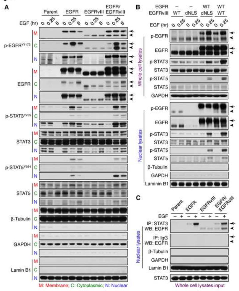
Figure 7.
EGFR and EGFRvIII cooperate to active STAT in the nucleus. (A) LN-229:parent, LN-229:EGFR, LN-229:EGFRvIII, or LN-229:EGFR/EGFRvIII cells were serum-starved for 24 hr then treated with EGF (50 ng/ml) for times shown. Samples were harvested, subject to subcellular fractionation to obtain membrane (M), cytoplasmic (C), and nuclear (N) extracts, and analyzed by immunoblot using antisera indicated. EGFR is indicated by arrow, whereas EGFRvIII is indicated by arrowhead. (B) LN-229:EGFRvIII, LN-229:EGFRvIIIdNLS, LN-229:EGFR/EGFRvIIIdNLS, or LN-229:EGFR/EGFRvIII were serum-starved for 24 hr then treated with EGF (50 ng/ml) for 0 and 15 min, whole cell lysates or nuclear extracts were analyzed by immunoblot using antisera indicated. (C) LN229:parent, LN-229:EGFR, LN-229:EGFRvIII, or LN-229:EGFR/EGFRvIII cells were serum-starved for 24 hr then treated with EGF (50 ng/ml) for 0 and 15 min, and fractionated to obtain nuclear extracts. Nuclear STAT3 was immunoprecipitated using a mouse monoclonal STAT3 antibody, and immunoprecipitates analyzed by immunoblot to detect EGFR and EGFRvIII (using a rabbit polyclonal EGFR antibody, which recognizes both EGFR and EGFRvIII). Efficacy of subcellular fractionation in A, B, and C is indicated by membrane and cytoplasmic marker protein β-Tubulin, cytoplasmic marker protein GAPDH, and nuclear marker protein Lamin B1. See also Figure S7.

