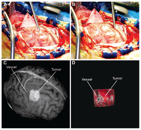
FIGURE 4.
Patient 3. A, intraoperative high-resolution image showing the surgical FOV with the tumor highlighted using forceps. B, intraoperative high-resolution image showing a significant vessel highlighted. Preoperative MR textured surface (C) and intraoperative textured LRS surface (D). The tumor and vessel highlighted in the digital photographs has been manually highlighted in each textured surface image.

