
Figure 3.
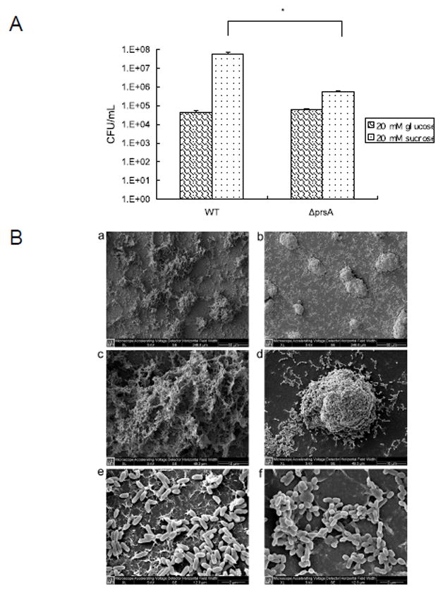
Biofilm formation characteristics. (A) Early Biofilm formation of S. mutans UA140 wild type and the prsA-deficient strains in minimal defined medium supplemented with glucose or sucrose. Each data point is the average of triplicate samples, and the error bars correspond to the standard deviations. The asterisk indicates that there was significantly less prsA mutant cells attached to the well of a 24-well flat-bottomed polystyrene microtiter plate than wild type in the presence of sucrose (Student’s t test p value <0.05). (B) Scanning electron micrographs of S. mutans 16 h-biofilms formed on glass surfaces. S. mutans UA140 wild-type biofilms (a, c, e); prsA-deficient strain biofilms (b, d, f). Magnifications, ×1000 (a, b), ×5000 (c, d) and ×20000 (e, f).