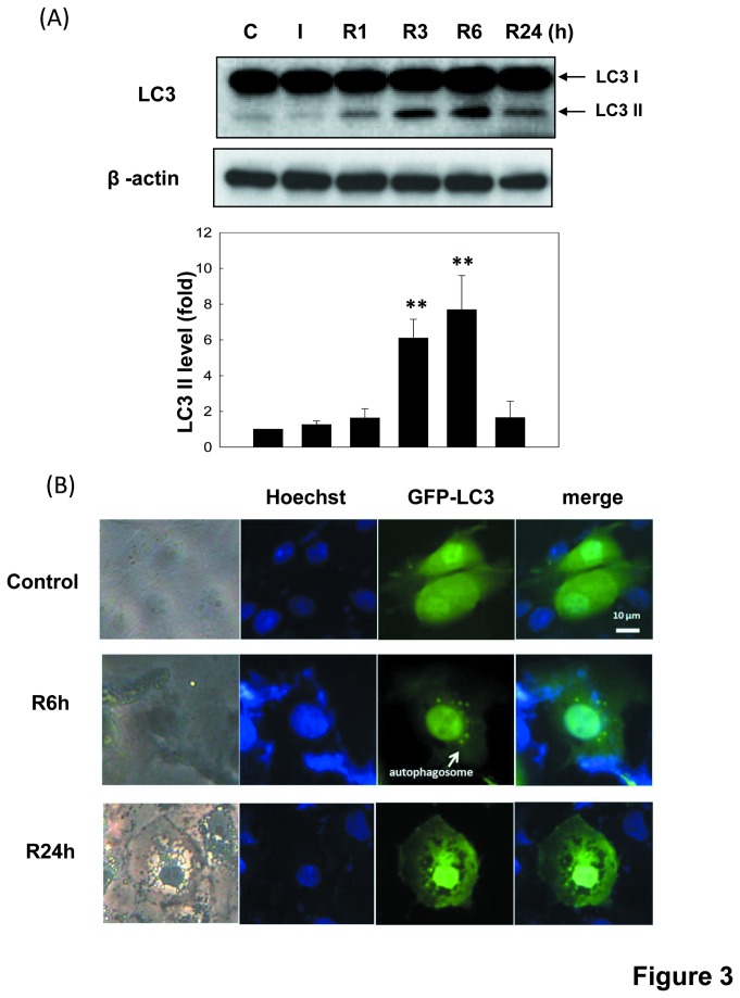
Figure 3. Autophagy in LLC-PK1 cells during in vitro I/R.
Cells were treated with 50 μM antimycin A and 5 mM 2-deoxyglucose for 1.5 h to induce ischemia (I) injury followed by reperfusion (R) for 1-24 h. In figure 3A, autophagy was determined by Western blotting using anti-LC3 antibody. The β-actin was used to an internal control. Data are presented as the means ± SDs in three independent experiments. *P < 0.05 and **P < 0.01 as compared with vehicle control group. In figure 3B, the GFP-LC3 puncta formation in LLC-PK1 cells was determined by immunofluorescence. Cells were transiently transfected with GFP-LC3 for 4 h before I/R treatment. Arrow indicates GFP-LC3 puncta formation (green). Nuclei were stained by Hoechst33258 dye (blue). Scale bar = 10 μm. Results shown are representative of at least three independent experiments.

