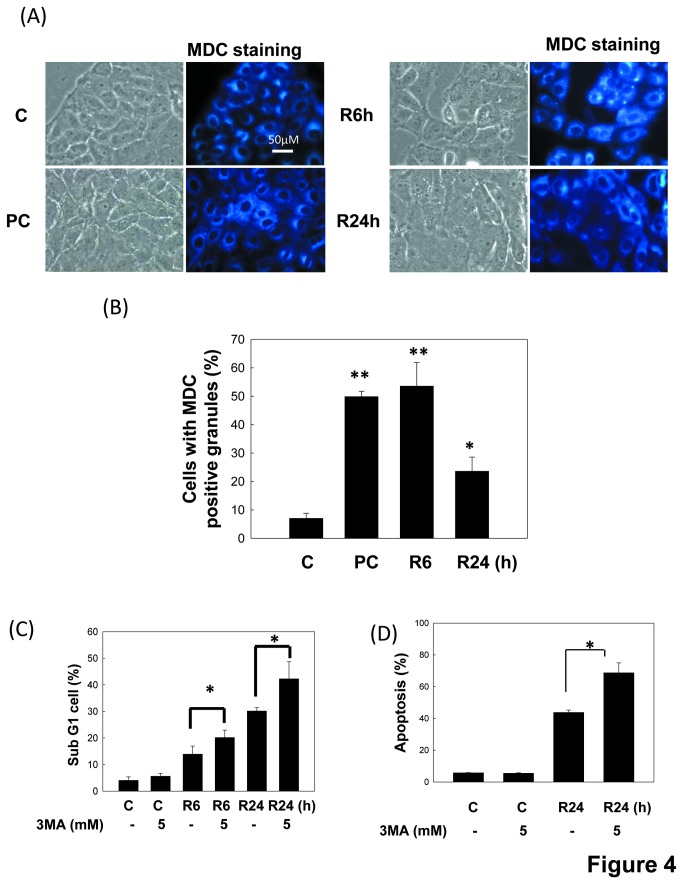
Figure 4. Enhancement of apoptosis by autophagy inhibitor in LLC-PK1 cells during in vitro I/R.
Cells were treated with 50 μM antimycin A and 5 mM 2-deoxyglucose for 1.5 h to induce ischemia (I) injury followed by reperfusion (R) for 6 or 24 h in the absence or presence of 5 mM 3MA. Cells were treated with 1 μM rapamycin for 6 h as an autophagic positive control. The MDC staining for autophagic vacuoles was examined by fluorescence microscopy (A). The number of MDC-positive cells was counted (a minimum of 100 cells per sample) (B). Moreover, the percentages of cells with the hypodiploid DNA content (sub-G1 cells) were determined by flow cytometry (C). Cell apoptosis was also performed by Annexin V and PI dual staining and determined by flow cytometry (D). In B, C and D, data are presented as the means ± SDs in three independent experiments. *P < 0.05 and **P < 0.01 as compared with vehicle control group. PC: positive control. C: control.

