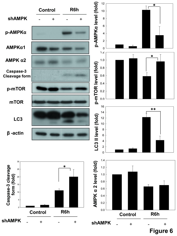
Figure 6. AMPK down-regulates the phosphorylation of mTOR and up-regulates the formation of LC3-II in LLC-PK1 cells during in vitro I/R.
Cells, which were transfected with scrambled shRNA (scra.) or shRNA for AMPKα (shAMPK), were treated with 50 μM antimycin A and 5 mM 2-deoxyglucose for 1.5 h to induce ischemia (I) injury followed by reperfusion (R) for 6 h. The protein expressions of phospho-AMPKα, phospho-mTOR, AMPKα1, AMPKα2, mTOR, caspase-3 and LC3 were determined by Western blotting. The β-actin was used to an internal control. Data are presented as the means ± SDs in three independent experiments. *P < 0.05 and **P < 0.01 as compared with vehicle control group.

