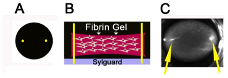
Figure 1. Schematic Diagram of 3-Dimensional Fibrin Gel.

A) Schematic top view of 24-well fibrin gel with pins shown in yellow. B). Schematic side-view of fibrin gels in 24-well plate. Fibrin gel containing primary fibroblasts is shown in pink. Yellow insect pins are inserted into a pre-plated layer of sylguard (shown in gray) to provide points of tension that enhance cell alignment and extracellular matrix deposition. C). Actual image of a fibrin gel containing cells cultured in a 24-well plate. Yellow arrows indicate insect pins.
