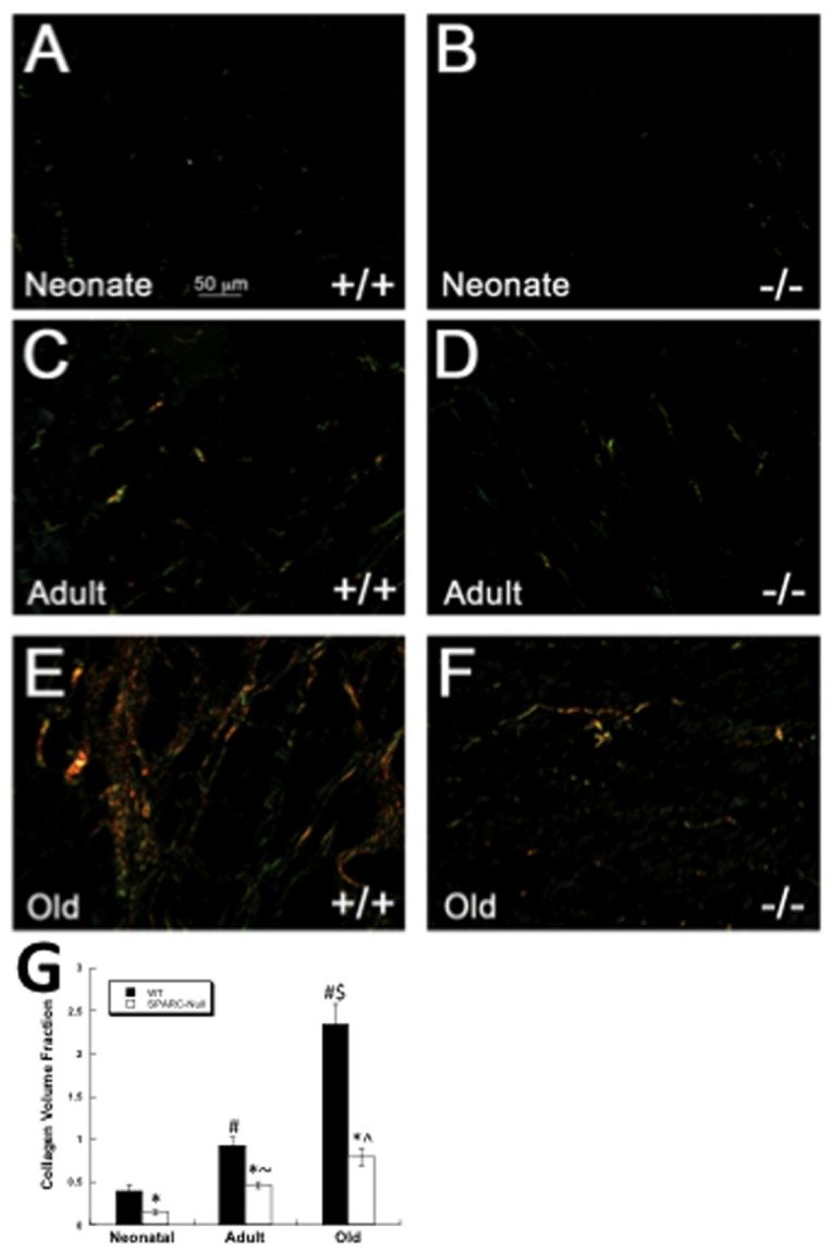
Figure 2. Cardiac Fibrillar Interstitial Collagen Changes with Age.

Picrosirius red stained images of cardiac sections viewed under polarized light. Sections from wild-type (WT, +/+) neonates (A) demonstrated diminished fibrillar collagen in comparison to WT adult (C) sections. Increased amounts of fibrillar collagen were apparent in hearts of WT old (E) mice. Cardiac sections taken from SPARC-null neonates (B, -/-), adult (D), and old (F) mice exhibited reduced amounts of PSR stained fibrillar collagen versus age-matched WT mice. Size bar in A = 50 µms; each panel is of equal magnification. G). Quantification of collagen volume fraction from neonate, adult and old hearts. Values presented for WT and SPARC-null adult and old animals were published previously [6]. * p<0.05 versus WT at each age, #p<0.05 vs. WT neonate, $ p<0.05 vs. WT adult, ^ p<0.05 vs. SP-null adult, ~p<0.05 vs. SP-null neonate.
